
(a) Retina
(b) Ear ossicles
(c) Cochlea
(d) Organ of Corti
(a) Retina:
The retina forms the finest internal, neurosensory layer of the eyeball. The surface of the retina interacts with the choroid and the interior interacts with the vitreous humor.
The outer surface has four layers:
Colored layer – Made up of a single layer of cells that contain a dark brown color.
Layers of photoreceptors – Consists of two types of cells – rods and tubes
Rods- Rod shaped and contains a reddish-brown protein known as rhodopsin or visible purple. Contains the content of vitamin A. Rods do not react with color and are sensitive to dim light. They offer vision in the dark, which is why it is known as evening vision or scotopic vision
Cones – Sensitive to colors and bright lights, providing daytime or visual imagery. The pigment found in lump cells is known as iodopsin. Three types of con cells respond to green, red, and blue light. Some colors are detected by a one-time trigger of more than one type of cell. White light sensation is performed when all three types of cells are triggered simultaneously.
Cone cells are not sensitive to low light and therefore in the dark, color cannot be seen.
(b) Ear Ossicles:
The middle ear contains three ossicles known as malleus, stapes and incus connected to each other by a chain pattern. The malleus is connected to the tympanic membrane and the stems are connected to the oval window of the cochlea. The ear ossicles increase the efficiency of the transmission of sound waves to the inner ear. The middle ear canal is connected to the pharynx using a Eustachian tube that helps balance pressure on both sides of the eardrum.
(c) Cochlea:
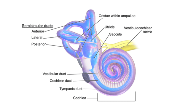
Cochlea is an integrated part of the labyrinth. The cochlea, basilar, and reissner’s membranes divide the perilymph covered larynx of the skeletal labyrinth into the upper scala vestibule and the lower scala tympanum. The scala media (space inside the cochlea) is full of endolymph and below the cochlea, the scala vestibule ends at the oval window and the scala tympani ends at a circular window opening in the middle ear.
(d) Organ of Corti:
Located on the basilar membrane of the inner ear, the cortium is an auditory organ. It contains hair cells with internal auditory receptor cells located in rows on the inner side of the organ. Stereo cilia are processes found in the apical edges of hair cells and the lower parts of hair cells contain synaptic contacts with associated nerve fibers. Just above the lines of these hair cells, a smooth layer of gelatinous known as the tectorial membrane is found.